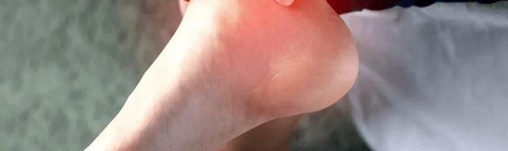
The pain showed up the way gout usually does: sudden, nasty, and out of proportion to anything you actually did. Except this time it isn’t your big toe. It’s the back of your heel, right above the shoe line, and going up a single stair makes you wince. So you search “heel pain,” read plantar fasciitis, grab a shoe insert, and two days later you can barely put weight on the foot.
Here is the part most pages skip: gout doesn’t only live in the big toe. The Achilles tendon, the thick cord running down the back of your heel, is one of the most common spots urate crystals build up outside the joints. If you’ve already dealt with gout in the ankle, this isn’t much of a leap. Let’s walk through what the imaging studies actually show, how to tell it apart from ordinary heel problems, and what to do about it.
If gout likes the big toe, why does it end up in the heel?
The Achilles tendon anchors your calf muscle to the heel bone. That anchor point, where tendon meets bone, is called an enthesis. It takes a pounding with every step you take, and researchers think that mix of constant mechanical strain and cooler tissue makes it an easy place for urate crystals to drop out and settle.
We have hard numbers on this. In a dual-energy CT study, a scan that colors urate crystals on imaging, researchers scanned both feet of 92 people with tophaceous gout. Crystals showed up in 10.8% of tendon and ligament sites overall, and the Achilles was the single most involved tendon, with crystals in 39.1% of all Achilles tendons examined. The peroneal tendons along the outside of the ankle came second at 18.1%. The plantar fascia on the sole of the foot was involved less than 5% of the time.
An ultrasound study out of New Zealand found much the same. Of 24 people with tophaceous gout, tophus material was visible inside 73% of their Achilles tendons (35 of 48 tendons scanned), and in none of the matched control group. Blood flow signals inside the tendon, a sign of active inflammation, were also far more common in the people with gout. A Chinese ultrasound study again ranked the Achilles first for urate deposits among the major leg tendons, ahead of the quadriceps and patellar tendons at the knee.
What does gout in the heel feel like?
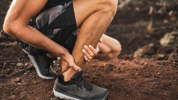
Most people describe pain at the back of the heel rather than the bottom of it. You might notice:
- Tenderness right where the tendon plugs into the heel bone, sometimes swelling a little way up the cord itself.
- Warmth and redness over the spot, with pain that arrives abruptly and peaks fast, the rhythm of a flare rather than a slow overuse ache.
- A firm little lump you can feel under the skin, which may be a tophus. More on those in our piece on tophi and how they’re managed.
- Stiffness after rest that loosens up a bit as you move around.
One detail trips people up. Because the Achilles is a tendon, not a joint, plenty of sufferers never call it gout. They assume they pulled something running. And yes, this can happen even in someone who has never been diagnosed before, since crystals can sit quietly in tendons for years before they cause trouble. When deposits show up in several spots at once, that picture is sometimes mistaken for gout moving across multiple joints.
Plantar fasciitis or gout? Where, exactly, does it hurt?
This is the question that sorts most cases. Location matters.
Plantar fasciitis hits the sole of the foot, classically under the inside of the heel, and the pain is often worst for the first few steps after you get out of bed. It builds gradually over weeks. Gout in the Achilles hurts at the back of the heel, often a little above the heel bone, comes on fast, and brings swelling and warmth with it. The plantar fascia, remember, is rarely involved in gout, showing deposits in fewer than 5% of sites in the CT study.
Heel pain as a whole is extremely common. A review in American Family Physician put it at about 2 million office visits a year in the United States. The same review noted that retrocalcaneal bursitis, inflammation of the small cushion between the tendon and heel bone, shows up most often in women aged 40 to 60 who have a bony prominence called a Haglund deformity. In active children and adolescents, Sever disease accounts for up to 16% of outpatient musculoskeletal conditions. All of these can overlap with gout in the same heel, which is why the pattern matters more than any single symptom.
How a clinician actually figures it out
Heel pain alone doesn’t answer the question. Imaging does.
Musculoskeletal ultrasound can show crystals in a few ways. The double-contour sign is a bright line of urate coating a cartilage surface, tophi appear as lumpy deposits, and aggregates show as bright speckles inside tendons. A meta-analysis of 13 studies found the double-contour sign had a specificity of 92% for gout, an ultrasound-visible tophus 94%, while sensitivity was more modest, around 66% and 56% for the two features taken one at a time. Put the findings together and sensitivity rose to about 80%. In plain terms: when ultrasound shows these signs, gout is quite likely, but a clean scan doesn’t fully rule it out.
Dual-energy CT is another option, and it is the tool that produced the 39.1% Achilles figure above. The 2015 gout classification criteria from the American College of Rheumatology and the European League Against Rheumatism accept either ultrasound double-contour or urate on dual-energy CT as imaging evidence, and those criteria carry a reported sensitivity of 92% and specificity of 89%. The gold standard stays the same as anywhere else in gout: drawing fluid and seeing urate crystals under a microscope.
What actually helps
Treat it like the flare it is. The usual options are a nonsteroidal anti-inflammatory drug (NSAID), colchicine, or a corticosteroid, chosen around your kidney function and other health issues, and they work best when started early. If this is new to you, our guide covers what to do in the first 12 hours of a flare. If you already take allopurinol or febuxostat, keep taking it through the attack. Stopping and restarting swings your urate level and tends to drag the pain out.
Long term, the answer is urate-lowering therapy taken to a target, generally below 6 mg/dL. Many rheumatologists aim lower, around 5 mg/dL, until visible tophi clear. That lower, steady level is what lets deposits in tendons dissolve back over months rather than merely quieting one episode.
Skip the aggressive deep massage and don’t dig at a lump yourself. A couple of symptoms deserve prompt attention rather than home care: fever or red streaks around the heel, a lump that breaks open and drains chalky material, or a pop you hear or feel followed by inability to push off the foot, which suggests a tendon rupture. The imaging studies offer some reassurance here. Even with crystals present, actual structural damage inside the tendon was uncommon in the ultrasound study, so finding gout in your Achilles doesn’t mean the tendon is on the verge of tearing.
Frequently Asked Questions
Q: Can gout really affect the Achilles tendon?
Yes. The Achilles is the most common tendon site for urate deposits in people with gout. Crystals were found in 39.1% of Achilles tendons in one CT study, and ultrasound found tophus material in 73% of tendons among people with tophaceous gout.
Q: How do I tell heel gout from plantar fasciitis?
By location and pace. Plantar fasciitis hurts on the sole under the heel, builds slowly, and is worst with the first morning steps. Gout in the Achilles hurts at the back of the heel, comes on fast, and usually brings swelling and warmth.
Q: Will urate crystals in the tendon dissolve?
With urate held consistently below target, deposits do gradually resolve, which is why medication adherence matters more than chasing each flare. Ultrasound studies suggest crystals can be present without much lasting structural damage, and keeping urate low is what tips that balance toward clearance.
Q: Ultrasound or dual-energy CT, which is better?
Ultrasound is quick, cheap, and repeatable, and the double-contour sign is quite specific. Dual-energy CT gives a detailed map of deposits and is useful when ultrasound is unclear or the diagnosis is in doubt. Your clinician picks based on what’s available and what the scan needs to answer.
Q: Could gout make my Achilles rupture?
It isn’t a common outcome. In the New Zealand ultrasound study, intratendinous structural damage was rare despite frequent crystal deposits. Most ruptures trace to other factors, like age, fluoroquinolone antibiotics, or steroid exposure. If you hear a pop and can’t push off the foot, that needs urgent evaluation.
Q: Should I stretch the tendon during a flare?
Gentle, comfortable movement is fine once the acute pain eases. Hold off on forceful calf stretches or heavy loading while the heel is hot and swollen, since pushing through it rarely shortens the flare.
References
- Dalbeth N, Kalluru R, Aati O, Horne A, Doyle AJ, McQueen FM. “Tendon involvement in the feet of patients with gout: a dual-energy CT study.” Annals of the Rheumatic Diseases. 2013 Sep;72(9):1545-1548. doi:10.1136/annrheumdis-2012-202786. PMID: 23334212
- Carroll M, Dalbeth N, Allen B, Stewart S, House T, Boocock M, Frampton C, Rome K. “Ultrasound Characteristics of the Achilles Tendon in Tophaceous Gout: A Comparison with Age- and Sex-matched Controls.” The Journal of Rheumatology. 2017 Oct;44(10):1487-1492. doi:10.3899/jrheum.170203. PMID: 28765249
- Deng SH, Wang J, Hu J, Tu B, Dang WT, Liu J. “Ultrasonographic Evaluation of Urate Crystal Deposition in Tendons With Gout Patients.” International Journal of Rheumatic Diseases. 2024 Dec;27(12):e70006. doi:10.1111/1756-185X.70006. PMID: 39676463
- Zhang Q, Gao F, Sun W, Ma J, Cheng L, Li Z. “The diagnostic performance of musculoskeletal ultrasound in gout: A systematic review and meta-analysis.” PLoS One. 2018 Jul 6;13(7):e0199672. doi:10.1371/journal.pone.0199672. PMID: 29979706
- Neogi T, Jansen TL, Dalbeth N, et al. “2015 Gout classification criteria: an American College of Rheumatology/European League Against Rheumatism collaborative initiative.” Annals of the Rheumatic Diseases. 2015 Oct;74(10):1789-1798. doi:10.1136/annrheumdis-2015-208237. PMID: 26359487
- FitzGerald JD, Dalbeth N, Mikuls T, et al. “2020 American College of Rheumatology Guideline for the Management of Gout.” Arthritis Care & Research. 2020 Jun;72(6):744-760. doi:10.1002/acr.24180. PMID: 32391934
- Morancie NA, Irvin L, Rayala BZ. “Heel Pain: Diagnosis and Management.” American Family Physician. 2025 Dec;112(6):648-656. PMID: 41533410
- Flores DV, Goes PK, Damer A, Huang BK. “The Heel Complex: Anatomy, Imaging, Pathologic Conditions, and Treatment.” Radiographics. 2024 Apr;44(4):e230163. doi:10.1148/rg.230163. PMID: 38512730
Reviewed by the GoutSavvy Editorial Team