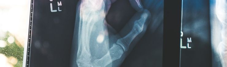
You notice a firm lump on your elbow. Or maybe it’s on the side of your big toe, or along a finger. It doesn’t hurt much at first, but it keeps growing. You might wonder what it is and whether it needs to come out.
That lump could be a tophus (plural: tophi), a deposit of uric acid crystals that builds up under the skin when gout goes untreated or poorly controlled for years. A lot of people assume tophi mean automatic surgery. They don’t. Most shrink with medication. But there are specific situations where a surgeon does need to get involved, and knowing the difference matters.
What Exactly Is a Tophus?
When your blood uric acid stays above about 6.8 mg/dL for long periods, the excess forms needle-shaped monosodium urate crystals. These crystals settle in joints and soft tissue. Your immune system attacks them, causing the fiery pain of a gout flare. But some crystals don’t trigger an acute attack. Instead, they quietly accumulate over months and years, walled off by inflammatory cells and fibrous tissue into a hard nodule. That’s a tophus.
Tophi usually show up after a decade or more of uncontrolled high uric acid, though they can appear sooner in severe cases. Common spots include the elbows, fingers, toes, ankles, knees, and even the outer ear. They range from pea-sized to lumps several centimeters across. Sometimes they break through the skin and drain a chalky white material, which can be alarming but is actually composed of urate crystals.
One review estimated that roughly 12 to 35 percent of people with gout develop visible tophi at some point, depending on how well their uric acid is managed. In parts of Southeast Asia where treatment access is limited, rates as high as 47 percent have been reported in referral center cohorts. That’s not a minor problem.
Medication Can Dissolve Tophi — Really
Here’s the part that surprises a lot of folks: the same urate-lowering drugs used to prevent flares can also make tophi shrink and disappear. The key is hitting and holding a lower target than you’d use for gout without tophi.
The 2020 American College of Rheumatology (ACR) guideline recommends starting urate-lowering therapy (ULT) for anyone with one or more subcutaneous tophi. For standard gout, the target serum urate is under 6 mg/dL. When tophi are present, rheumatologists usually aim for under 5 mg/dL until the deposits fully resolve. That lower target speeds up crystal dissolution considerably.
The evidence backs this up. The NOR-Gout study, a five-year treat-to-target trial from Norway published in Annals of the Rheumatic Diseases in 2026, followed 209 patients on ULT. At the five-year mark, 71.2 percent had uric acid below 360 µmol/L (roughly 6 mg/dL), and ultrasound showed complete tophi resolution in 63.2 percent of patients who had them at baseline. Only 16 percent reported a flare in the previous year, and those who did had higher uric acid levels.
How long does it take? It depends on the size of the tophus and how low your uric acid goes. A prospective Chinese study using ultrasound to track tophi found significant size reduction within six months of treat-to-target ULT, with continued shrinkage through 12 months. For larger deposits, the process can take years. Patience matters here, which is why sticking with medication is so important, even when you feel fine.
For refractory cases where oral drugs don’t lower uric acid enough, pegloticase (Krystexxa), an infused enzyme that breaks down uric acid directly, can drop serum urate below 1 mg/dL. A 2019 analysis of two phase III trials found that among responders, tophi shrank at an average rate of about 60 mm² per month, with projected complete resolution in roughly 10 months. That said, pegloticase is reserved for severe, treatment-resistant gout because of cost, infusion reactions, and the need for biweekly IV treatment.
If you’re already on ULT but still developing tophi, it’s worth asking your doctor whether your dose is high enough or whether adding a second agent makes sense. You can read more about why medication sometimes falls short in our piece on why your gout medication isn’t working.
When Surgery Actually Becomes Necessary
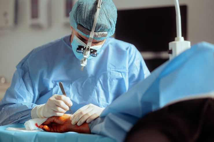
Surgery for tophi is the exception, not the rule. A 2025 retrospective study of 28 patients in PLoS ONE found that surgery was most often performed to restore or improve joint function, not just to remove a lump. The researchers identified specific scenarios where surgical intervention was justified:
Nerve compression. Tophi can press on nerves, causing numbness, tingling, or weakness. The most common example is carpal tunnel syndrome in the wrist, where a tophus squeezes the median nerve. About 5 to 10 percent of people with tophaceous gout develop carpal tunnel symptoms. Surgery involves releasing the compressed ligament and removing the urate deposits around the nerve.
Tendon involvement or rupture. When urate crystals infiltrate a tendon, they weaken it. The tendon can fray or even snap. This is particularly serious in the hands (extensor tendons that let you straighten your fingers) and the Achilles tendon at the back of the heel. A ruptured tendon needs surgical repair, and the tophus contributing to the damage gets cleaned out at the same time. We covered this in more detail in our article about Achilles tendon rupture as a hidden gout danger.
Skin ulceration and infection. Sometimes a tophus erodes through the skin, creating an open wound that drains chalky material. If it gets infected, that’s a medical emergency. An infected tophus behaves like a complicated skin and soft tissue infection. It needs urgent surgical debridement and antibiotics, usually covering Staphylococcus aureus including MRSA. A 2016 review in Seminars in Arthritis and Rheumatism noted that infected or ulcerated tophi have much higher wound complication rates, with delayed healing reported in up to two-thirds of cases in older series.
Mechanical obstruction. A large tophus can physically block a joint from moving, make it impossible to wear shoes, or interfere with basic daily activities. If medication hasn’t reduced it enough and it’s genuinely disabling, surgical removal is reasonable.
Diagnostic uncertainty. Occasionally a soft tissue mass looks like a tophus but could be something else, such as a rheumatoid nodule or even a tumor. If imaging and aspiration can’t confirm the diagnosis, a surgeon may remove it for examination under a microscope.
Cosmetically bothersome tophi alone are generally not considered a strong reason for surgery, though some surgeons will remove them electively if they cause significant distress. The risk of wound complications usually outweighs the cosmetic benefit for small, asymptomatic deposits.
What the Surgery Involves
The approach depends on where the tophus is and how deeply it has infiltrated surrounding tissue.
Open excision is the traditional method. The surgeon makes an incision over the tophus and removes the chalky urate material, carefully scraping it off tendons, nerves, and joint surfaces. This works well for large, complex deposits but involves a longer incision and more tissue disruption.
Arthroscopic or endoscopic removal is less invasive. Using small portals and a camera, the surgeon debrides tophaceous material from inside or around a joint with motorized instruments. A 2026 case report in Cureus described endoscopic removal of an ankle tophus through three tiny incisions, with no postoperative infection or skin necrosis. Recovery tends to be faster, though the technique requires specialized training and isn’t suitable for every location.
Joint reconstruction may be needed when tophi have destroyed bone and cartilage. Options include joint fusion (arthrodesis) for severely damaged joints, or occasionally joint replacement. These are major procedures reserved for advanced cases where function has been lost.
The Risks Are Real
Tophus surgery isn’t risk-free, and the complication rates run higher than for routine soft tissue surgery. Data from hand surgery series show:
- Wound breakdown or delayed healing: roughly 10 to 20 percent. Urate deposits can interfere with normal tissue healing, and skin over a large tophus is often thin and stretched.
- Infection: 5 to 10 percent, higher when the tophus was already draining or ulcerated.
- Tendon injury: around 5 percent, since crystals can be glued to tendon fibers and separating them is delicate work.
- Nerve injury: under 5 percent, but possible when tophi wrap around nerves.
- Recurrence: 5 to 10 percent, almost always in people who don’t maintain ULT after surgery.
The 2025 PLoS ONE study reported better-than-average results, with 97 percent of surgical sites healing primarily (65 of 67 sites) and only minimal complications. The authors credited careful patient selection and operating during disease remission when inflammatory markers were low. They found that patients with elevated C-reactive protein (CRP), erythrocyte sedimentation rate (ESR), or white blood cell counts had significantly longer hospital stays, supporting the idea that timing matters. Surgery during an active flare is generally avoided unless there’s an infection that can’t wait.
After Surgery, You Still Need Medication
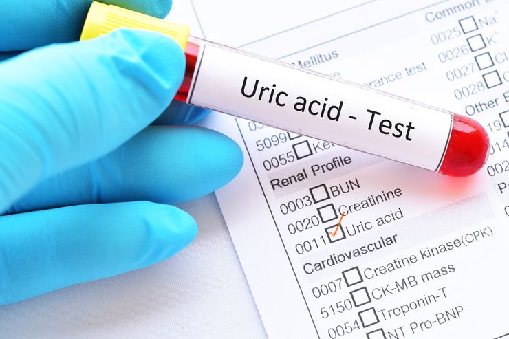
This is the part nobody likes to hear but everyone needs to understand: surgery removes the visible deposits, not the underlying problem. Your body is still producing too much uric acid or not excreting enough of it. If you stop ULT after surgery, tophi come back.
The 2025 retrospective study drove this point home. Among 22 patients who stayed on ULT after surgery, gout flares and new tophi both dropped significantly compared to the six patients who didn’t continue medication. Surgery provided a short-term urate-lowering effect simply by removing a large urate reservoir, but the long-term benefit depended largely on continued medical therapy.
After surgery, your doctor will likely aim for a serum urate target below 6 mg/dL, or below 5 mg/dL if any tophi remain. You may also take a low-dose anti-inflammatory, such as colchicine 0.6 mg daily, for the first few months to prevent flares triggered by rapid urate shifts as crystals mobilize. This is the same prophylaxis recommended whenever ULT is started, and you can learn more about the importance of consistent treatment in our coverage of the gout medication adherence crisis.
Why Early Treatment Prevents All This
The simplest way to deal with tophi is to never get them in the first place. Once crystals start accumulating, every year of uncontrolled uric acid adds to the burden. The deposits can erode bone, destroy cartilage, weaken tendons, and permanently damage joints. Tophaceous gout is also associated with higher mortality and reduced quality of life compared to gout without tophi.
If you’ve had a gout flare, talk to your doctor about checking your serum urate and discussing whether ULT makes sense. The old idea that you should wait for a certain number of flares before starting medication is outdated. Current ACR guidelines recommend ULT after even two flares, and immediately if you have tophi, chronic kidney disease, or a history of uric acid kidney stones. For more on kidney-related complications, see our article about the gout-ckd treatment gap.
Questions People Often Ask
Can tophi go away on their own without medication?
It’s extremely unlikely. Tophi are solid deposits of urate crystals that don’t dissolve unless serum urate drops well below the saturation threshold of roughly 6.8 mg/dL. Dietary changes alone rarely lower uric acid enough to resolve established tophi. Urate-lowering medication is almost always necessary.
How long does it take for a tophus to shrink with medication?
It depends on the size and how low your uric acid goes. Small tophi may start shrinking within three to six months. Larger deposits can take one to several years. The NOR-Gout study found that 63 percent of patients had complete ultrasound-detected tophi resolution after five years of treat-to-target ULT. The lower your uric acid (consistently under 5 mg/dL), the faster the process.
Is tophus removal painful?
The surgery itself is done under anesthesia, so you won’t feel it during the procedure. Postoperative discomfort varies depending on the location and extent of dissection. Your surgical team will prescribe pain management, and most people report significant relief once the wound heals, especially if the tophus had been causing pressure or nerve symptoms.
Can I drain a tophus at home if it’s leaking?
No. If a tophus breaks through the skin, keep the area clean with mild soap and water, cover it with a sterile dressing, and contact your doctor promptly. Draining or squeezing it yourself can introduce bacteria and cause a serious infection. Watch for spreading redness, warmth, fever, or increasing pain, which could signal infection and require urgent medical care.
Does insurance cover tophus surgery?
Most insurance plans cover tophus removal when there’s a documented medical reason, such as nerve compression, tendon damage, infection, or significant functional impairment. Purely cosmetic removal may not be covered. Check with your insurer and ask your surgeon’s office to help with preauthorization if needed.
What’s the difference between a tophus and a gout flare lump?
A gout flare causes temporary swelling from inflammation that usually subsides within days to a couple of weeks. A tophus is a permanent deposit of crystals that stays and grows over months or years. Tophi are typically firm to the touch and not as red or hot as an acutely inflamed joint, though they can become inflamed during a flare.
References
- Hammer HB, Karoliussen L, Terslev L, et al. “Ultrasound-detected crystal depositions and clinical flares dissolve during successful urate-lowering therapy: 5-year follow-up results from the treat-to-target NOR-Gout study.” Annals of the Rheumatic Diseases. 2026;85(2):379-387. doi:10.1016/j.ard.2025.10.029.
- Zhang T, Yang B, Xu X, et al. “Evaluation of surgical treatment of gout: A retrospective study on 28 cases with tophi.” PLoS ONE. 2025;20(1):e0313586. doi:10.1371/journal.pone.0313586.
- Mandell BF, Yeo AE, Lipsky PE. “Tophus resolution in patients with chronic refractory gout who have persistent urate-lowering responses to pegloticase.” Arthritis Research & Therapy. 2018;20:286. doi:10.1186/s13075-018-1782-4.
- FitzGerald JD, Dalbeth N, Mikuls T, et al. “2020 American College of Rheumatology guideline for the management of gout.” Arthritis Care & Research. 2020;72(6):744-760. doi:10.1002/acr.24180.
- Wang J, Xu Y, Yun X, et al. “Research progress and clinical application of surgical treatment for tophaceous gout.” Journal of Clinical Personalized Medicine. 2026;5(3):195-201. doi:10.12677/jcpm.2026.53199.
- Kasper IR, Juriga MD, Giurini JM, Shmerling RH. “Treatment of tophaceous gout: When medication is not enough.” Seminars in Arthritis and Rheumatism. 2016;45(6):669-678. doi:10.1016/j.semarthrit.2015.10.006.
- Liu Y, Ma Q, Zhang Y, et al. “Musculoskeletal ultrasound in monitoring the efficacy of gout: A prospective study based on tophus and double contour sign.” Balkan Medical Journal. 2023;40(2):126-132. doi:10.4274/balkanmedj.galenos.2022.2022-7-34.
- Chhana A, Lee G, Dalbeth N. “Factors influencing the crystallization of monosodium urate: a systematic literature review.” BMC Musculoskeletal Disorders. 2015;16:296. doi:10.1186/s12891-015-0752-7.
Reviewed by the GoutSavvy Editorial Team