Getting an accurate gout diagnosis is the first step toward effective treatment. But diagnosing gout isn’t always straightforward. Many conditions cause joint pain and swelling, and gout can mimic other disorders. Here’s what you need to know about how doctors diagnose gout.
Understanding the Diagnostic Challenge
Gout is the most common form of inflammatory arthritis in the United States and United Kingdom, yet diagnosing it can be tricky. The classic presentation—a sudden, extremely painful flare affecting the big toe—doesn’t occur in most people. Some people have atypical symptoms that could point to several different conditions.
The Clinical Assessment
Medical History
Your doctor will start by taking a detailed medical history, asking about:
- Current symptoms and their onset
- Previous similar episodes
- Family history of gout or arthritis
- Dietary habits and alcohol consumption
- Medications you’re taking
- Other medical conditions (especially kidney disease, diabetes, or hypertension)
Physical Examination
During the physical exam, your doctor will:
- Examine the affected joint(s) for signs of inflammation
- Check for tophi (hard deposits under the skin)
- Assess the pattern of joint involvement
- Look for signs of chronic disease
Laboratory Tests
Serum Uric Acid Test
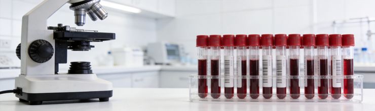
The serum uric acid (SUA) test measures the amount of uric acid in your blood. This is typically the first laboratory test ordered when gout is suspected. For tips on lowering your uric acid levels through diet, see our comprehensive guide.
Important points about SUA testing:
- Hyperuricemia (high uric acid levels) (SUA above 6.8 mg/dL or 400 µmol/L) is present in about 95% of people with gout
- However, a normal SUA level does NOT rule out gout
- SUA levels can actually DROP during a gout flare due to inflammation
- The best time to measure SUA is 2 weeks or more after a flare subsides
- Multiple tests may be needed to establish your baseline level
According to MedlinePlus, a uric acid blood test may be used to help diagnose gout, usually in combination with synovial fluid analysis, and to monitor uric acid levels during cancer treatment.
Joint Fluid Analysis (Arthrocentesis)
Joint fluid analysis is considered the gold standard for gout diagnosis. During this procedure, a doctor uses a needle to withdraw fluid from the affected joint. If you’re experiencing recurrent gout flares, discuss whether joint aspiration might help confirm your diagnosis.
The fluid is then examined under a polarized microscope to look for monosodium urate (uric acid crystals) (MSU) crystals. In people with gout, these crystals appear as:
- Needle-shaped crystals
- Yellow when aligned parallel to the slow axis of a red compensator
- Blue when aligned perpendicular to the compensator (negative birefringence)
According to Medscape, needle-shaped (visible under a microscope) urate crystals are seen in approximately 85% of synovial fluid specimens from people with gout.
Why Joint Aspiration Is Rarely Done in Primary Care
Despite being the most definitive test, joint aspiration is rarely performed in primary care settings because:
- Samples should be tested quickly and protected from light
- Most practices only collect samples once daily
- Results can be unreliable if timing isn’t optimal
- The procedure requires specialized equipment and training
Joint aspiration is more commonly performed in specialist rheumatology settings when the diagnosis remains uncertain.
Other Blood Tests
Your doctor may order additional tests to assess:
- Kidney function (creatinine, estimated glomerular filtration rate (eGFR))
- Complete blood count
- Inflammatory markers (erythrocyte sedimentation rate (ESR), C-reactive protein (CRP))
- Blood glucose
- Lipid levels
Imaging Studies
X-Rays
X-rays are not useful for detecting early gout because urate deposits aren’t visible on standard X-rays. However, they can show:
- Chronic joint damage from repeated flares
- Bone erosions characteristic of long-standing gout
- Rule out other conditions like fractures or osteoarthritis
Ultrasound
Musculoskeletal ultrasound is becoming increasingly useful for gout diagnosis. Key findings include:
- Double contour sign: Urate deposits on the cartilage surface
- Snowstorm appearance: Floating particles in synovial fluid
- Tophi visualization
- Synovial inflammation
Ultrasound has the advantage of being non-invasive, readily available, and able to detect early crystal deposition.
Dual-Energy CT (DECT)
Dual-energy CT (DECT) is a specialized imaging technique that can detect urate deposits anywhere in the body, including in joints that haven’t yet become symptomatic. According to UK guidelines, DECT has good sensitivity and specificity to confirm or exclude gout diagnosis.
However, DECT availability is limited in many clinical settings, and its cost makes it impractical for routine use.
The 2015 American College of Rheumatology (ACR)/European Alliance of Associations for Rheumatology (EULAR) Classification Criteria
In 2015, the American College of Rheumatology (ACR) and the European Alliance of Associations for Rheumatology (EULAR) established updated classification criteria for gout. These criteria are primarily used in research but can guide clinical diagnosis.
Entry Criterion
For these criteria to apply, you must have at least one episode of swelling, pain, and tenderness in a peripheral joint or bursa.
Scoring System
Points are assigned based on clinical, laboratory, and imaging findings:
Clinical Features
- Pattern of joint involvement (ankle/midfoot = 1 point; first metatarsophalangeal (MTP) joint = 2 points)
- Characteristics of flares (one feature = 1 point, all three features = 2 points)
- Tophi present = 4 points
Laboratory Findings
- SUA level (4 to <6 mg/dL = 2 points; 6 to <8 mg/dL = 3 points; 8 to <10 mg/dL = 4 points; ≥10 mg/dL = 4 points)
Imaging
- Double contour sign on ultrasound = 4 points
- Urate deposits on DECT = 4 points
Definite Diagnosis
A score of 8 or higher confirms gout classification. The presence of MSU crystals in joint fluid or tophus provides a definitive diagnosis regardless of score. Understanding your diagnosis can help you better manage your condition. Consider reading about how purines affect your body and potential gout complications.
The Role of Tophi in Diagnosis
Tophi are deposits of urate crystals that can form under the skin, typically around joints, ears, or finger pads. While tophi usually develop in patients with long-standing gout, their presence is highly specific for gout diagnosis.
Tophi may appear as:
- Painless lumps under the skin
- White or yellowish deposits
- Chalky texture
- Varying sizes from microscopic to several centimeters
If you notice any unusual lumps or nodules near your joints, mention this to your doctor, as tophi can aid in diagnosis even without a confirmed gout flare history.
Preparing for Your Doctor Visit
Being prepared for your appointment can help ensure an accurate diagnosis. Consider the following tips:
What to Bring
- A list of all your symptoms, including when they started and how long they lasted
- Photos of any visible joint swelling or skin changes
- A complete list of all medications and supplements you take
- Information about previous gout attacks if any
- A family health history, particularly for gout or arthritis
Questions to Ask Your Doctor
- What tests do I need to confirm gout?
- What are my uric acid levels, and what do they mean?
- What treatment options are available?
- How can I prevent future attacks?
- Should I see a specialist?
Keeping a Symptom Diary
Before your appointment, try to document:
- When symptoms started
- What you were doing when symptoms began
- Any foods or drinks consumed before the attack
- Any medications started recently
- How long the symptoms lasted
For more information on managing gout after diagnosis, see our comprehensive guide to gout-friendly eating.
Understanding Your Lab Results
What Is Hyperuricemia?
Hyperuricemia is defined as having serum uric acid levels above 6.8 mg/dL (400 µmol/L), which is the approximate limit of urate solubility in the blood. However, it’s important to understand that:
- Hyperuricemia is necessary but not sufficient for gout diagnosis
- Most people with hyperuricemia never develop gout
- Some people with gout have normal SUA levels during or between flares
Interpreting Your Results
Work with your healthcare provider to understand your specific lab results in context. Factors that affect interpretation include:
- When the test was performed relative to your last flare
- Your kidney function
- Your medications
- Other medical conditions
- Diet and alcohol intake around the time of testing
When Diagnosis Is Uncertain
If your doctor is uncertain about the diagnosis, they may refer you to a rheumatologist for:
- Joint aspiration with microscopy
- Advanced imaging
- Evaluation of other conditions that can mimic gout
Differential Diagnoses
Conditions that can look like gout include:
- Pseudogout (calcium pyrophosphate deposition (CPPD) disease) – calcium pyrophosphate crystals
- Septic arthritis – infection in the joint
- Rheumatoid arthritis
- Psoriatic arthritis
- Cellulitis
- Reactive arthritis
Frequently Asked Questions
What blood test confirms gout?
No single blood test definitively confirms gout. Serum uric acid testing is helpful but can be normal during an acute attack. The gold standard for diagnosis is joint fluid analysis showing urate crystals under polarized microscopy. This test can confirm gout with certainty.
Can imaging detect gout?
Yes. Ultrasound can show characteristic double-contour sign and snowstorm appearance indicating urate crystals. Dual-energy CT scan can specifically identify urate deposits and is useful for detecting early or hidden gout. X-rays typically only show gout changes in advanced disease.
Why is proper gout diagnosis important?
Misdiagnosis is common because gout symptoms resemble other conditions like pseudogout, infection, or rheumatoid arthritis. Proper diagnosis ensures you receive appropriate treatment and avoids unnecessary medications for conditions you do not have.
When should I see a specialist for gout diagnosis?
A rheumatologist is the specialist best equipped to diagnose and manage gout. Consider referral if diagnosis is uncertain, flares are frequent, you have kidney disease, or initial treatments are not controlling your condition adequately.
References
- Gout: Diagnosis and Management. British Society for Rheumatology, June 2022. https://www.ncbi.nlm.nih.gov/books/NBK583528/
- Gout. StatPearls – NCBI Bookshelf, 2025. https://www.ncbi.nlm.nih.gov/books/NBK546606/
- Gout and Pseudogout Workup. Medscape, 2025. https://emedicine.medscape.com/article/329958-workup
- Uric Acid Test. MedlinePlus. https://medlineplus.gov/lab-tests/uric-acid-test/
- Chen-Xu M, Yokose C, Rai SK, Pillinger MH, Choi HK. Contemporary Prevalence of Gout and Hyperuricemia in the United States. Arthritis Rheumatol. 2019;71(5):764-770. PubMed
- American College of Rheumatology. 2020 Guideline for the Management of Gout. Arthritis Care & Research. 2020. PubMed
Reviewed by the GoutSavvy Editorial Team