Your joints feel fine. No pain, no swelling, nothing. Maybe you even forgot you have gout.
Then another attack hits, and the cycle repeats.
Here’s what most people don’t realize: that pain-free stretch between flares isn’t healing time. It’s damage time.
The Deceptive Quiet Period
Picture this: You wake up, stretch your ankles, flex your fingers. Everything moves smoothly. No warnings, no hints that anything’s wrong. You might even think, “Maybe my gout isn’t that serious after all.”
You’re not alone in thinking this. It’s exactly what your doctor sees too: they take X-rays early on and the images often look completely normal. That validation feels good. Maybe you can skip that uric acid pill today.
Except you can’t.
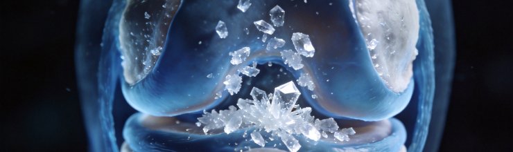
The truth is, monosodium urate (MSU) crystals, the microscopic culprits behind every gout flare, are still there. Still accumulating. Still doing damage you can’t feel.
What Your Joints Look Like During the “Quiet” Period
Crystal Accumulation
When your serum uric acid stays above 6.8 mg/dL (the point where crystals start forming). MSU crystals deposit throughout your body. These aren’t just hanging out harmlessly, they’re embedding themselves in:
- The big toe joint (where most attacks start), but also ankles, knees, wrists
- Tendons and ligaments (explaining why some people develop Achilles tendinitis)
- Bursae, the fluid-filled sacs that cushion your joints
- Soft tissues around joints
- Your kidneys (where they contribute to stones and declining kidney function)
Think of it like calcium deposits in pipes. You don’t notice anything until the water flow slows down. Except with gout, you might not notice until a pipe bursts.
The Constant Inflammatory Battle
Even without obvious flares, your immune system has not given up. White blood cells are continuously trying to clear those crystals. This isn’t the dramatic, painful inflammation of a flare, it’s a low-grade, chronic inflammatory response that:
- Thickens and damages the synovium (your joint lining)
- Erodes surrounding cartilage
- Gradually destroys bone
- Creates bone cysts filled with crystal deposits
Inside Tophi
Most people know tophi as those visible lumps that pop up on fingers or ears. But the real damage happens before you can see anything:
- Microscopic tophi form deep within joint structures
- They erode cartilage from the inside out
- Bone destruction spreads beneath cartilage
- By the time you see a visible tophus, significant damage has already occurred
Here’s the concerning part: these lumps don’t just appear suddenly. They grow slowly over years, often silently. If you’ve had elevated uric acid for 5, 10, or 15 years without treatment, there’s a good chance some crystal deposition has occurred even if you’ve never noticed a visible lump.
When tophi do become visible, they signal substantial internal damage. The visible portion is like the tip of an iceberg. What you cannot see beneath the skin is usually much larger.
Imaging: Seeing What’s Actually Happening
Traditional X-rays miss most of this early damage. That’s where modern imaging comes in:
Ultrasound
A Doppler ultrasound can reveal:
- Double contour sign, crystal coating on cartilage surface
- Snowstorm appearance, crystals swirling in synovial fluid
- Tophi before they become clinically visible
- Active inflammation in the joint lining
The good news? Ultrasound is non-invasive, uses no radiation, and can be done in a regular clinic appointment.
MRI
Magnetic resonance imaging goes deeper:
- Bone marrow edema, often visible before actual erosions appear
- Soft tissue tophi embedded in tissues
- Tendon and ligament involvement
- Chronic synovitis (long-term joint lining inflammation)
Dual-Energy CT (DECT)
This specialized scan is becoming the gold standard for detecting urate deposits:
- Color-codes urate crystals (making them impossible to miss)
- Quantifies total crystal burden
- Detects deposits anywhere in the body
- Monitors changes over time with treatment
Studies using dual-energy CT (DECT) have been sobering. Patients with “asymptomatic hyperuricemia”, elevated uric acid but no flares, have frequently shown significant crystal deposits. A study published in the Annals of the Rheumatic Diseases found that over 20% of people with elevated uric acid but no history of gout attacks had visible tophi on ultrasound.
Think about that: One in five people walking around with elevated uric acid already have crystal deposits, and many do not know it.
The takeaway from imaging studies isn’t meant to scare you. It’s meant to show you why “feeling fine” isn’t the same as “being fine.” If you know there’s damage happening, you can do something about it before it becomes irreversible.
The Kidney Angle Nobody Talks About
Between flares, your kidneys are taking hits too:
- Urate crystals deposit in kidney tissue
- Chronic interstitial nephritis develops slowly
- Blood pressure tends to increase
- Kidney function gradually declines
Here’s the vicious part: Kidney damage impairs uric acid excretion, raising your uric acid levels, which causes more kidney damage. Without intervention, this cycle accelerates.
The connection between gout and kidney disease is often overlooked because kidney problems do not announce themselves early. You might not feel any different until significant function is lost. That’s why regular kidney monitoring, through blood tests measuring creatinine and estimated glomerular filtration rate (eGFR), is essential for anyone with gout.
Why Treatment Matters, Even When You Feel Fine
The goal of urate-lowering therapy isn’t just preventing the next painful attack. It’s preventing permanent damage:
- Bone erosions rarely reverse. Once bone is eaten away, it doesn’t typically grow back.
- Large tophi cause lasting problems. They can damage tendons, cause joint deformity, and sometimes require surgery to remove.
- Cartilage loss is permanent. Unlike some tissues, cartilage has limited ability to repair itself.
- Early intervention gives the best results. Less damage accumulated means more function preserved.
Some patients worry about taking medication long-term. That’s understandable. But consider this: the most common urate-lowering drugs (like allopurinol and febuxostat) have been used for decades. The risks of untreated gout, chronic pain, joint destruction, kidney damage, typically outweigh the risks of treatment for most patients.
Your doctor can help you weigh the pros and cons based on your specific situation.
The Numbers Don’t Lie
Long-term studies paint a clear picture:
- Joint erosion progresses during asymptomatic periods, yes, even when you’re feeling fine
- Each flare adds damage, but damage continues between flares too
- Undertreated patients (uric acid still above target) accumulate more damage
- Aggressive urate-lowering therapy can actually reduce crystal burden over time
What’s Your Target?
Current guidelines recommend:
- All patients with visible tophi should receive urate-lowering therapy
- Anyone with frequent flares (2 or more per year) should be treated
- Some patients benefit from lower targets (uric acid below 5 mg/dL)
- Treatment is typically lifelong, “drug holidays” allow damage to resume
The key metric: your serum uric acid level. If you’re on medication, ask your doctor what your current level is and what the target should be. Numbers matter here.
What You Can Do
If you’re between flares and feeling fine, here’s what actually matters:
- Don’t skip your uric acid medication because you feel okay
- Ask about advanced imaging (ultrasound or DECT) to assess your actual crystal burden
- Get your kidneys checked. Blood tests and urine analysis can detect early damage
- Know your numbers: serum uric acid, kidney function
- Don’t assume diet alone will solve the problem, while cherries and certain foods can help, most people with gout need medication to reach target uric acid levels
The gap between “I feel fine” and “my joints are fine” can be enormous. Modern imaging has shown us just how much damage can accumulate silently. The good news: this damage is often preventable, and sometimes even reversible, with proper treatment.
Frequently Asked Questions
Can joint damage from gout be reversed?
Some damage can improve with aggressive urate-lowering therapy. Tophi can shrink, inflammation resolves, and some bone density may recover. However, established bone erosions and cartilage loss are largely permanent, making early treatment critical rather than optional.
How often should I have imaging to check for damage?
It depends on your situation. Patients with frequent flares or visible tophi may benefit from annual ultrasound assessment. Discuss with your rheumatologist what monitoring schedule makes sense for your specific case, there isn’t one answer for everyone.
I only have one flare a year. Do I really need daily medication?
Even infrequent flares cause cumulative damage. Current guidelines increasingly support treating tophi and frequent flares, but the decision depends on your uric acid level, imaging evidence of damage, kidney function, and personal risk factors. This is worth a detailed discussion with your doctor.
Will my gout eventually burn itself out?
No. While flare frequency sometimes decreases over time, gout is a progressive disease. Without treatment, crystal burden typically increases, and damage continues. Most patients experience continued progression as they age.
How do I know if damage is happening even when I feel fine?
Advanced imaging, particularly ultrasound and DECT, can detect urate deposits and joint damage before symptoms appear. If you’re concerned about your crystal burden, ask your rheumatologist whether advanced imaging makes sense for you.
References
- FitzGerald JD, et al. “2020 American College of Rheumatology Guideline for the Management of Gout.” Arthritis Care & Research. 2020;72(6):744-760. PubMed
- Ogdie A, Taylor WJ. “Imaging of gout: an atlas.” Current Opinion in Rheumatology. 2025;37(2):73-85. LWW
- De Miguel E, Puig JG, Castillo C, et al. “Diagnosis of gout in patients with asymptomatic hyperuricaemia: a pilot ultrasound study.” Annals of the Rheumatic Diseases. 2012;71(2):157-158. PubMed
- Dalbeth N, et al. “Urate crystal deposition and bone erosion in gout: ‘inside-out’ or ‘outside-in’? A dual-energy computed tomography study.” Arthritis Research & Therapy. 2016;18:188. PMC
- Choi HK, et al. “Clinical utility of dual energy computed tomography in gout: current concepts and applications.” Acta Bio-Medica Atenei Parmensis. 2020;91(Suppl 11):1-9. PMC
Reviewed by the GoutSavvy Editorial Team